
Plastinates are resin-preserved body parts used in anatomical teaching
Medical students to use plastinated human tissue made famous in Body Worlds exhibition
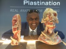
The University of Exeter Medical School has acquired ‘plastinates’ – resin-preserved body parts – famously used in the Body Worlds exhibit for use in teaching anatomy.
The University of Exeter Medical School has acquired ‘plastinates’ – resin preserved body parts – famously used in the Body Worlds exhibit for use in teaching anatomy.
The plastinates include two full body slices (male and female), hearts, brains, two upper limbs, a lower limb dissected from pelvis to toes, a chest , kidneys and half a face.
They are far more practical for use in teaching anatomy as these are non-toxic and, unlike cadavers, will last 15-20 years. The preserved human tissue in total cost £60,000.
These resources are praised for their anatomical accuracy and visibility. Students are able to better appreciate details which would otherwise be hard to demonstrate in a classroom environment.
Vikram Devaraj, Senior Lecturer and Clinical Lead, said: “People were stunned when they were displayed in exhibitions, and now they are a superb teaching resource. They are real human beings, allowing relationships of organs and structures to be seen. It helps students to see, for example, why a shoulder will dislocate forward and therefore how to put one back when they diagnose one!
"The full body slices also display the precise location, depth and paths taken by nerves and vessels, revealing their vulnerability to trauma and a surgeon’s knife.
"This allows for accurate demonstrations of clinical procedures, ways to stay safe when practising as a doctor and an understanding of the location of vitally important structures”
Plastination is a process used to preserve bodies or body parts for use in anatomy invented by Dr Gunther von Hagens in 1977. Water and fat in the body are replaced by certain plastics to create specimens which can be touched, do not decay and retain most of their original properties.
The Body Worlds exhibition opened in 1996 and has toured across Europe, Asia and America. With over 200 anatomical specimens, the exhibit continues to shock audiences.
These plastinates will be used in the entirely new core curriculum to be introduced for current first year BMBS students, along with the following additions and changes:
- New core curriculum for 2018 Year 1 BMBS Program
- Major revision of content and sequence of study - focusing on human morphology by systems (e.g. musculoskeletal, neuro, endocrine, cardiac, respiratory)
- New visuals for ELE with enhanced newly embedded interactive content
- Study Guides extensively rewritten; now clearer with additional signposting to e-resources for computer aided learning (CAL)
- New case studies stripped back to basics to emphasize the core concepts discussed in PBL
- New medical imaging content to support anatomy learning, written within a smaller team: improved content, closely linked to previous material
- Animal organ dissection workshops including heart, brain and kidneys
- Alioscopy High Definition display screens to display anatomical features in 3 Dimensions
- All core areas in anatomy and physiology (essential for the clinical pathway weeks in years 3 and 4) embedded on the ELE platform, to guide early years learning
- Online Video master classes available in a range of topics. Some themed with anatomy and physiology e.g. wound healing and the management and treatment of burns.
Date: 3 October 2018