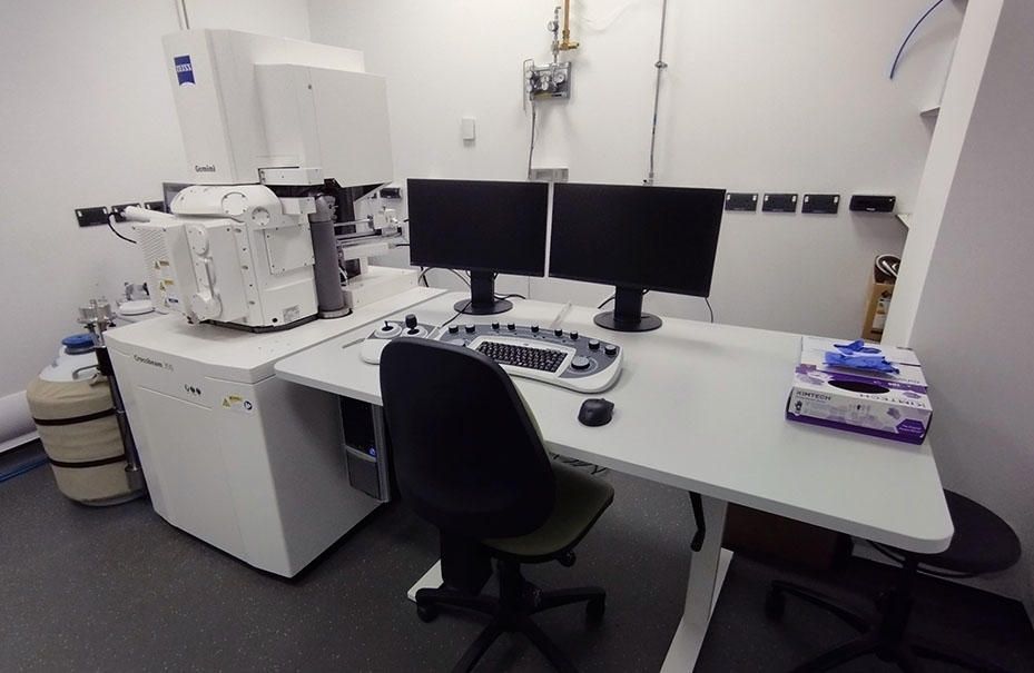
Zeiss Crossbeam 350 cryo-SEM
The Zeiss Crossbeam 350 Cryo-SEM combines advanced field-emission scanning electron microscopy with dedicated cryogenic sample preparation and transfer capabilities, enabling high-resolution imaging of hydrated, soft and temperature-sensitive specimens in their near-native state. Equipped with a Quorum PP3010T cryo preparation and transfer system, the platform supports a broad range of cryo-SEM applications, including biological tissues, cells, plant material, biofilms, hydrogels, emulsions, nanoparticles and other hydrated or semi-liquid samples.
The system offers exceptional flexibility through a range of specialist cryogenic specimen holders designed to accommodate diverse sample types. These include a standard cryo holder compatible with multiple specimen stubs and rivets for the rapid freezing and imaging of liquids and semi-liquid samples, an extended-length shuttle for larger specimens, and a dedicated leaf-clamping shuttle optimised for plant and environmental samples. Together, these accessories enable researchers to preserve delicate structures and investigate samples that would be difficult or impossible to analyse using conventional electron microscopy methods.
For imaging and analytical applications, the Crossbeam 350 is equipped with multiple complementary detector systems. Secondary electron imaging is provided by the SE2 detector for topographical contrast, while the InLens secondary electron detector delivers high-resolution surface imaging at low accelerating voltages. Compositional and material contrast can be obtained using both the InLens Energy Selective Backscatter (ESB) detector and a six-sector annular solid-state backscatter detector (BSD), supporting a wide range of structural and analytical workflows. Instrument operation and advanced image acquisition are controlled through ZEISS SmartSEM, providing an intuitive environment for routine imaging as well as advanced microscopy applications.
Key Features
- Dedicated Quorum PP3010T Cryo-SEM preparation and transfer system
- Multiple cryogenic specimen holders including standard, extended-length and leaf-clamping shuttles
- Imaging of hydrated, soft, frozen and temperature-sensitive samples
- SE2 secondary electron detector for topographic imaging
- High-resolution InLens secondary electron detector
- InLens ESB detector for compositional and backscattered electron imaging
- Six-sector annular solid-state BSE detector
- Variable pressure operation for challenging and non-conductive samples
- SmartSEM software environment for microscope control and image acquisition
- Compatible with a broad range of biological, environmental and materials science applications
Accelerating voltage: between 0.01 to 30 kV
Applications
- Cryo-SEM imaging of hydrated biological specimens
- Plant and environmental sample analysis
- Biofilm and microbial imaging
- Soft matter research
- Nanoparticle and biomaterial characterisation
- Freeze-fracture studies
- Correlative cryogenic workflows
- High-resolution surface morphology analysis
- Imaging of non-conductive and difficult-to-prepare specimens
This platform provides Exeter researchers with a highly versatile cryogenic electron microscopy capability, bridging the gap between conventional SEM and advanced cryogenic imaging workflows while preserving samples in a state that closely reflects their native structure.