Living Systems Institute.
A unique scientific community focussed on the fundamental rules of life
We combine theory and technology from mathematical and physical sciences with experimental analysis in biology and biomedicine. The fusing of disciplines empowers research discovery across scales; from atom to organism, from microbe to human. Our shared vision is to decode the complex machinery of living systems and harness the underlying mechanisms to benefit health and wellbeing.
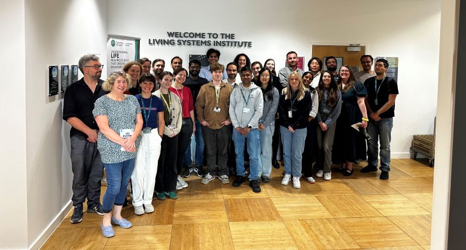
LSI Community
The LSI is a community of some 200 talented PhD students, post-doctoral researchers and principal investigators from around the world supported by excellent professional services staff. We collaborate to generate exciting discoveries and create new knowledge. Diversity is a major strength that enriches our science and our culture. Our vibrant and inclusive research environment is underpinned by dynamic PhD student and post-doc networks.
Latest news
See all news